This website is intended for Healthcare Professionals practicing in the U.S.

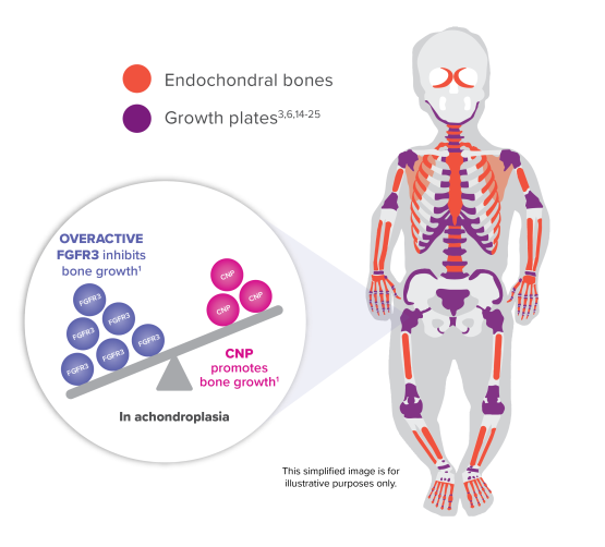
In achondroplasia, endochondral bone growth is inhibited throughout the body1-4

Endochondral bone growth, in which cartilage is replaced by bone at open growth plates, requires a balance of cell signals—CNP (which promotes bone growth) and FGFR3 (which slows bone growth).1,3

Endochondral bones make up >90% of the bones in the body4-13

Overactive FGFR3 signaling relative to CNP signaling in growth plate cells is the underlying cause of inhibited bone growth in achondroplasia. Endogenous CNP levels cannot adequately regulate overactive FGFR3 signals.1

Learn more about inhibited bone growth and the impact of timely care in achondroplasia.1,2

Download Brochure

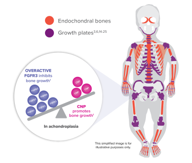
Infographic showing FGFR3 inhibiting endochondral bone growth and CNP promoting growth in growth plates in achondroplasia

CNP, C-type natriuretic peptide; FGFR3, fibroblast growth factor receptor 3.

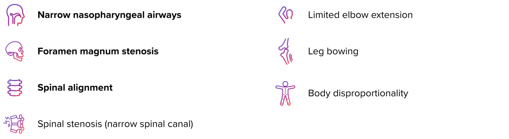
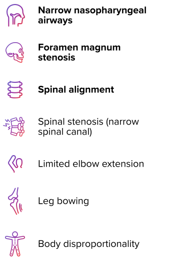
Achondroplasia is associated with potential multisystemic skeletal complications26

Many complications are more common in the first 2 years of life.2,26-28

image
iamge

VOXZOGO has been approved to increase linear growth in children with achondroplasia and open growth plates.29 Proactive and multidisciplinary care can help inform management and treatment approaches, which are important for multisystemic complications.2,30

Inhibited bone growth throughout the body can lead to reduced and disproportionate growth1-4,27

Disproportionate growth (differences in body proportions) is a characteristic feature of achondroplasia associated with reduced mobility, pain, and dependence on devices and caregiver assistance.27,31

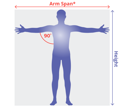
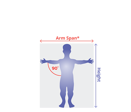
Achondroplasia affects how body proportions change over time, which is commonly measured by arm span, arm span-to-height ratio, and upper-to-lower body segment ratio.31-34

Arm span in adults with achondroplasia is ~35% shorter than average stature31

Arm span-to-height ratio: Arm span divided by standing height34

  • In average-stature individuals, arm span is shorter than height before puberty and greater than height after mid-puberty34
  • In achondroplasia, arm span is shorter than height before and after puberty (deficit increasing with age)31,34
arm
body-proportionality
Average Stature
Achondroplasia

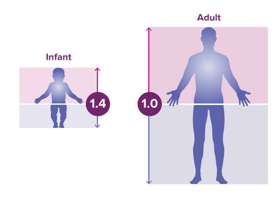
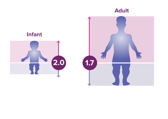
Upper body remains longer than the lower body in adults with achondroplasia32,33

Upper-to-lower body segment ratio: Length of the upper body divided by the length of the lower body.34

  • In average-stature individuals, upper body length at birth is greater than lower body length (ratio >1) but becomes proportional at 10 years old (ratio=1)32,33
  • In achondroplasia, upper body length at birth is greater than lower body length (ratio >1) but remains disproportionate into adulthood (ratio >1)32,33
Comparison infographic of average stature vs achondroplasia showing upper-to-lower body segment ratios from infancy to adulthood
Average Stature
Achondroplasia

*Length between tips of the left and right middle fingers when standing against a flat wall with arms outstretched, creating a 90-degree angle with the torso.34
Ratios represent the 50th percentile of children with achondroplasia.32,33

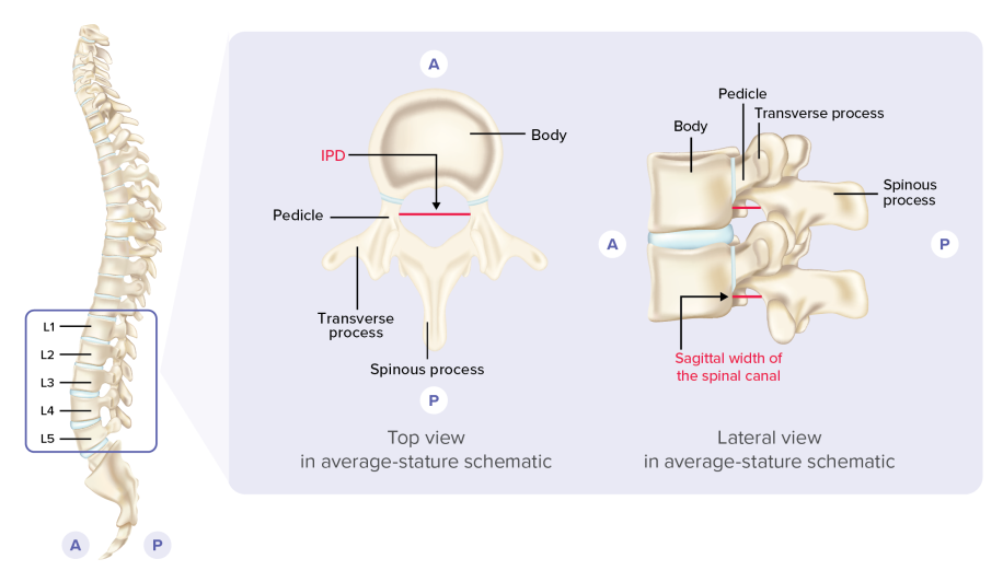
Achondroplasia is associated with the development of spinal stenosis26

Spinal stenosis (narrowing of the spinal canal that compresses the spinal cord/nerve roots) in the lumbar spine is often symptomatic in adulthood, associated with chronic pain, walking intolerance, bowel/bladder dysfunction, and paraplegia.26,35,36

However, its development can begin much earlier in children with achondroplasia, given that their spinal canal reaches ~95% of adult size before 5 years old.35,36

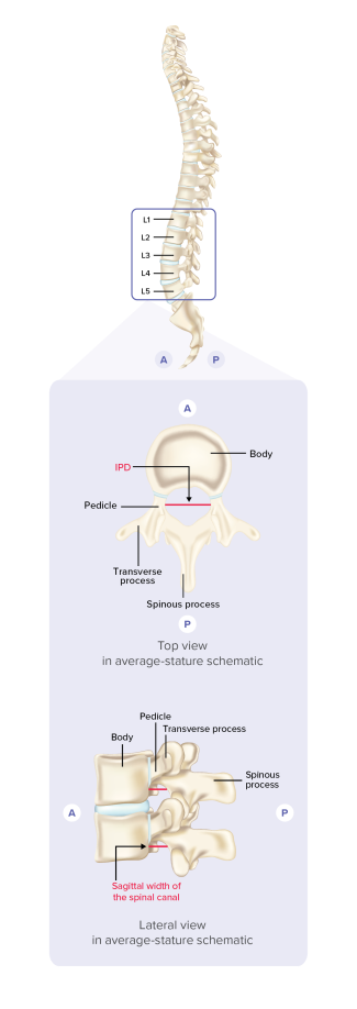
Common spinal canal measures related to spinal stenosis35:

  • Narrowing of interpedicular distance (IPD) on A/P X-ray
  • Reduced sagittal width of the spinal canal on lateral X-ray
Depiction of common spinal canal measures related to spinal stenosis in achondroplasia

A, anterior (front); L1-5, lumbar vertebra 1-5; P, posterior (back).  

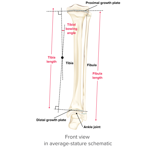
Leg bowing is a common orthopedic complication of achondroplasia37

In achondroplasia, leg bowing is associated with knee joint instability and skeletal irregularities of the lower leg, including tibial bowing and fibula overgrowth.37

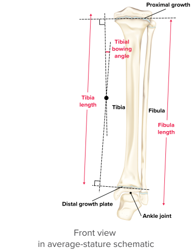
Lower leg bone morphology X-ray measures37:

  • Tibial bowing angle >0°, measured by linear intersections derived from the perpendicular of the proximal and distal growth plates of the tibia
  • Increased fibula/tibia (F/T) ratio (fibula length [cm]/tibia length [cm]) as a marker of overall fibula overgrowth relative to tibia growth
Depiction of lower leg bone morphology measures related to leg bowing in achondroplasia

Maximize time on treatment by starting early3,29,38

Learn more about efficacy and ongoing studies.

References:

  1. Horton WA, Hall JG, Hecht JT. Achondroplasia. Lancet. 2007;370(9582):162-172.
  2. Savarirayan R, Ireland P, Irving M, et al. International Consensus Statement on the diagnosis, multidisciplinary management and lifelong care of individuals with achondroplasia. Nat Rev Endocrinol. 2022;18(3):173-189.
  3. Mackie EJ, Tatarczuch L, Mirams M. The skeleton: a multi-functional complex organ: the growth plate chondrocyte and endochondral ossification. J Endocrinol. 2011;211(2):109-121.
  4. Clarke B. Normal bone anatomy and physiology. Clin J Am Soc Nephrol. 2008;3(suppl 3):S131-S139.
  5. Breeland G, Sinkler MA, Menezes RG. Embryology, bone ossification. In: StatPearls. StatPearls Publishing; 2023. Accessed March 17, 2026. https://www.ncbi.nlm.nih.gov/books/NBK539718/
  6. Berendsen AD, Olsen BR. Bone development. Bone. 2015;80:14-18.
  7. Cowan PT, Launico MV, Kahai P. Anatomy, bones. In: StatPearls. StatPearls Publishing; 2024. Accessed March 17, 2026. https://www.ncbi.nlm.nih.gov/books/NBK537199/
  8. Johns Hopkins Medicine. Anatomy of the bone. Accessed March 17, 2026. https://www.hopkinsmedicine.org/health/wellness-and-prevention/anatomy-of-the-bone
  9. Jin SW, Sim KB, Kim SD. Development and growth of the normal cranial vault: an embryologic review. J Korean Neurosurg Soc. 2016;59(3):192-196.
  10. Anderson BW, Kortz MW, Black AC, et al. Anatomy, head and neck, skull. In: StatPearls. StatPearls Publishing; 2023. Accessed March 17, 2026. https://www.ncbi.nlm.nih.gov/books/NBK499834/
  11. Encyclopaedia Britannica. Science & Tech. Skull. Accessed March 17, 2026. https://www.britannica.com/science/skull
  12. Hall R, Beals K, Neumann H, et al. Introduction to Human Osteology. Grand Valley State University; 2008.
  13. Encyclopaedia Britannica. Science & Tech. Clavicle. Accessed March 17, 2026. https://www.britannica.com/science/clavicle
  14. Baron J, Sävendahl L, De Luca F, et al. Short and tall stature: a new paradigm emerges. Nat Rev Endocrinol. 2015;11(12):735-746.
  15. Shahzad F. Pediatric mandible reconstruction: controversies and considerations. Plast Reconstr Surg Glob Open. 2020;8(12):e3285.
  16. Bartleby. Henry Gray (1825-1861). Anatomy of the Human Body. 1918. Fig. 237. Accessed March 17, 2026. https://www.bartleby.com/lit-hub/anatomy-of-the-human-body/fig-237/
  17. Encyclopaedia Britannica. Science & Tech. Pelvis. Accessed March 17, 2026. https://www.britannica.com/science/pelvis
  18. Mayo Clinic. Growth plate fracture. Accessed March 17, 2026. https://www.mayoclinic.org/diseases-conditions/growth-plate-fractures/multimedia/growth-plate-fracture/img-20005879
  19. International Center for Limb Lengthening. Growth arrest. Accessed March 17, 2026. https://www.limblength.org/conditions/growth-arrest/
  20. Hsieh YL, Wei X, Wang Y, et al. Chondrocyte Tsc1 controls cranial base bone development by restraining the premature differentiation of synchondroses. Bone. 2021;153:116142.
  21. Musculoskeletal Key. Cranial and pelvic “vertebrae” are they real vertebrae? Accessed March 17, 2026. https://musculoskeletalkey.com/cranial-and-pelvic-vertebrae-are-they-real-vertebrae/
  22. Young M, Selleri L, Capellini TD. Genetics of scapula and pelvis development: an evolutionary perspective. Curr Top Dev Biol. 2019;132:311-349.
  23. Yu M, Wang SM. Anatomy, head and neck, ethmoid bone. In: StatPearls. StatPearls Publishing; 2025. Accessed March 17, 2026. https://www.ncbi.nlm.nih.gov/books/NBK544328/
  24. Çankaya H, Egeli E, Kutluhan A, Kiriş M. Pneumatization of the concha inferior as a cause of nasal obstruction. Rhinology. 2001;39(2):109-111.
  25. Mitomo K, Matsunaga S, Kitamura K, et al. Sphenoid bone hypoplasia is a skeletal phenotype of cleidocranial dysplasia in a mouse model and patients. Bone. 2019;120:176-186.
  26. Hoover-Fong J, Cheung MS, Fano V, et al. Lifetime impact of achondroplasia: current evidence and perspectives on the natural history. Bone. 2021;146:115872.
  27. Witt S, Rohenkohl A, Bullinger M, et al. Understanding, assessing and improving health-related quality of life of young people with achondroplasia – a collaboration between a patient organization and academic medicine. Pediatr Endocrinol Rev. 2017;15(suppl 1):109-118.
  28. Brinkmann G, Schlitt H, Zorowka P, Spranger J. Cognitive skills in achondroplasia. Am J Med Genet. 1993;47(5):800-804.
  29. VOXZOGO [package insert]. Novato, CA: BioMarin Pharmaceutical Inc; 2024.
  30. Hoover-Fong J, Scott CI, Jones MC; Committee on Genetics. Health supervision for people with achondroplasia. Pediatrics. 2020;145(6):e20201010.
  31. Merker A, Neumeyer L, Hertel NT, et al. Development of body proportions in achondroplasia: sitting height, leg length, arm span, and foot length. Am J Med Genet A. 2018;176(9):1819-1829.
  32. Hoover-Fong JE, Schulze KJ, McGready J, et al. Age-appropriate body mass index in children with achondroplasia: interpretation in relation to indexes of height. Am J Clin Nutr. 2008;88(2):364-371.
  33. Chilbule SK, Dutt V, Madhuri V. Limb lengthening in achondroplasia. Indian J Orthop. 2016;50(4):397-405.
  34. Nwosu BU, Lee MM. Evaluation of short and tall stature in children. Am Fam Physician. 2008;78(5):597-604.
  35. Irving M, Savarirayan R, Hoover-Fong JE, et al. Effect of vosoritide on spine morphology in children  with achondroplasia: 1-year results from a randomized phase 2 study. J Endocr Soc. 2026;10(3):bvag008.
  36. Savarirayan R, Irving M, Maixner W, et al. Rationale, design, and methods of a randomized, controlled, open-label clinical trial with open-label extension to investigate the safety of vosoritide in infants, and young children with achondroplasia at risk of requiring cervicomedullary decompression surgery. Sci Prog. 2021;104(1):368504211003782.
  37. White KK, Irving M, Mukherjee S, et al. Effect of vosoritide on genu varum in children with achondroplasia after 1 year in randomized placebo-controlled trials. J Endocr Soc. 2026;10(3):bvag024.
  38. Savarirayan R, Hoover-Fong J, Ozono K, et al. International consensus guidelines on the implementation and monitoring of vosoritide therapy in individuals with achondroplasia. Nat Rev Endocrinol. 2025;21(5):314-324.
Contact Us

INDICATION AND IMPORTANT SAFETY INFORMATION

Warnings and Precautions for Risk of Low Blood Pressure
Transient decreases in blood pressure were observed in clinical studies. Patients with significant cardiac or vascular disease and patients on anti-hypertensive medicinal products were excluded from participation in VOXZOGO clinical trials. To reduce the risk of a decrease in blood pressure and associated symptoms (dizziness, fatigue, and/or nausea), patients should be well hydrated, have adequate food intake, and drink approximately 8-10 ounces of fluid in the hour prior to VOXZOGO administration.

In a 52-week, randomized, double-blind, placebo-controlled trial in 121 subjects with achondroplasia, subjects aged from 5.1 to 14.9 years, (Study 1) eight (13%) of 60 patients treated with VOXZOGO had a total of 11 events of transient decrease in blood pressure, compared to 3 (5%) of 61 patients on placebo, over a 52-week treatment period. The median time to onset from injection was 31 (18 to 120) minutes, with resolution within 31 (5 to 90) minutes in VOXZOGO-treated subjects. Two out of 60 (3%) VOXZOGO-treated patients each had one symptomatic episode of decreased blood pressure with vomiting and/or dizziness compared to 0 of 61 (0%) patients on placebo.

Adverse Reactions:
Adverse reactions that occurred in ≥5% of patients treated with VOXZOGO and at a rate greater than that of placebo in the phase 3 study are injection site reactions (including erythema, swelling, urticaria, pain, bruising, pruritus, hemorrhage, discoloration, and induration), vomiting, arthralgia, decrease in blood pressure, gastroenteritis, diarrhea, dizziness, ear pain, influenza, fatigue, seasonal allergy, and dry skin. VOXZOGO-treated patients had an increase in alkaline phosphatase levels (17%), and was noted as a laboratory abnormality.

Injection site reactions: In Study 1, injection site reactions occurred in 51 (85%) subjects receiving VOXZOGO and 50 (82%) subjects receiving placebo over a 52-week period of treatment. Patients receiving VOXZOGO experienced a total of 6983 events of injection site reactions, while patients receiving placebo experienced a total of 1776 events of injection site reactions, over a 52-week period, representing 120.4 events per patient/year exposure and 29.2 events per patient/year exposure, respectively. Two patients in the VOXZOGO arm discontinued treatment due to adverse events of pain and anxiety with injections.

Pediatric Patients 0 to <5 Years:
The safety of VOXZOGO in pediatric patients 0 to <5 years with achondroplasia was evaluated in a 52-week randomized, double-blind, placebo-controlled study (Study 2). In this study, 64 patients from birth to <5 years of age were randomized to receive either a daily vosoritide dose with similar exposure to that characterized to be safe and effective in children with ACH aged ≥5 years old, or placebo. An additional 11 patients received open-label treatment as part of this study. The most common adverse reactions (>10%) reported in pediatric patients 0 to <5 years were injection site reactions (86%) and rash (28%). The overall safety profile of VOXZOGO in pediatric patients 0 to <5 years was similar to that seen in older pediatric patients.

Administration and Monitoring:
VOXZOGO is administered as a daily subcutaneous injection. Prior to use, instruct caregivers on proper preparation and administration of VOXZOGO, and ensure caregivers have demonstrated the ability to perform a subcutaneous injection.

Monitor and assess patient body weight, growth, and physical development regularly every 3-6 months. Adjust dosage according to the patient’s actual body weight. Permanently discontinue treatment with VOXZOGO upon confirmation of no further growth potential, indicated by closure of epiphyses.

Special Populations:

  • There are no available data on the use of VOXZOGO in pregnant women, or data on the presence of VOXZOGO in human milk, the effects on the breastfed infant, or the effects on milk production.
  • The influence of renal impairment on the pharmacokinetics of VOXZOGO has not been evaluated. No dosage adjustment is needed for patients with eGFR ≥60 mL/min/1.73 m2. VOXZOGO is not recommended for patients with eGFR <60 mL/min/1.73 m2.

You may report side effects to the FDA at 1-800-FDA-1088 or www.fda.gov/medwatch. You may also report side effects to BioMarin at 1-866-906-6100.

Please see additional safety information in the full Prescribing Information.

VOXZOGO® (vosoritide) is indicated to increase linear growth in pediatric patients with achondroplasia and open growth plates.

  • This indication is approved under accelerated approval based on an improvement in annualized growth velocity. Continued approval for this indication may be contingent upon verification and description of clinical benefit in confirmatory trial(s).