

Though blindness occurs later in the progression of CLN2 disease, visual abnormalities may be identified using certain ophthalmologic assessments.1
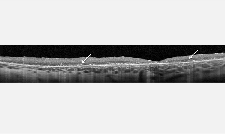
OCT can demonstrate progression of CLN2 disease via retinal degeneration and accumulation of hyper-reflective material.2,3
Note: This image is of an 8-year-old patient, reflecting late-stage advances of CLN2 disease.2

FA can be used to evaluate vascular leakage.2
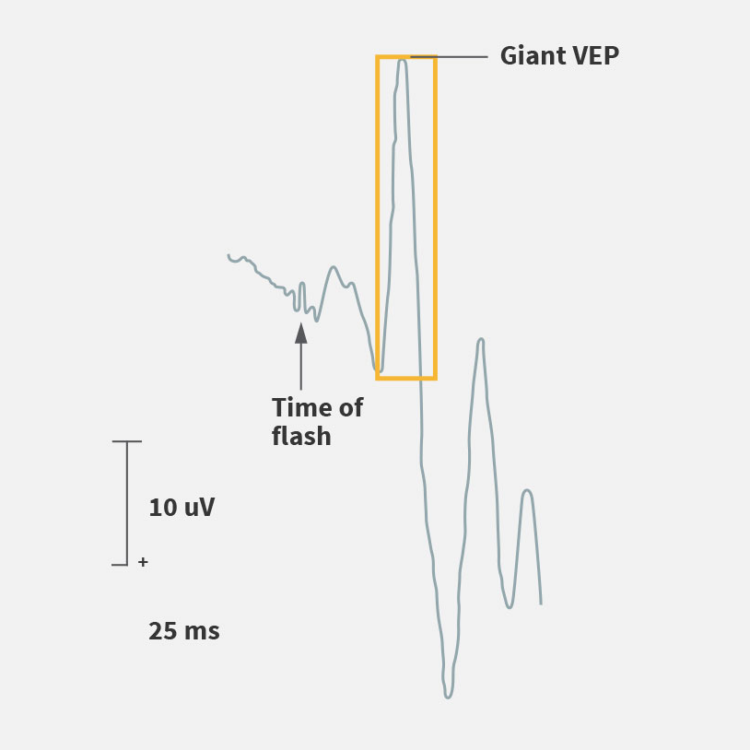
VEPs are abnormally enhanced until late in the disease and diminish in the final stage of the disease.4
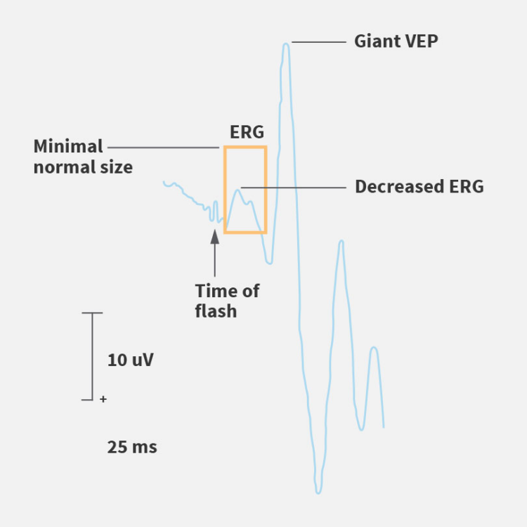
ERG may be diminished before visual deterioration is clinically detected.4,5

Electron microscopy (EM)
Although the use of electron microscopy in clinical practice has decreased worldwide, it is used to detect the distinct curvilinear structure of ceroid lipofuscin in suspected cases, often when other tests are inconclusive.1