This website is intended for Healthcare Professionals in Europe, Canada, Australia and New Zealand only.

What is the cause of achondroplasia?

A mutation of the FGFR3 gene

Achondroplasia is the most common type of skeletal dysplasia

Achondroplasia occurs in 1 out of 25,000 live births and affects around 250,000 people worldwide1,2 Achondroplasia accounts for nearly 90% of disproportionate short stature or dwarfism. Characterized by impaired endochondral bone growth, it is caused by a gain-of-function pathogenic variant in the fibroblast growth factor receptor 3 (FGFR3) gene and has distinct physical characteristics1,3-8

Silhouette of a child with achondroplasia highlighting five key physical characteristics

Physical characteristics, such as height, are indicators of bone growth throughout the body.

FGFR3 affects endochondral bone growth throughout the entire body9

Endochondral ossification, the replacement of cartilage by bone, occurs throughout the body and is involved in the development of roughly 90% of all bones. This process begins in utero and continues into early adulthood.10-12

Understanding endochondral bone growth at every level

In endochondral ossification, the precursor to prospective bone is cartilage

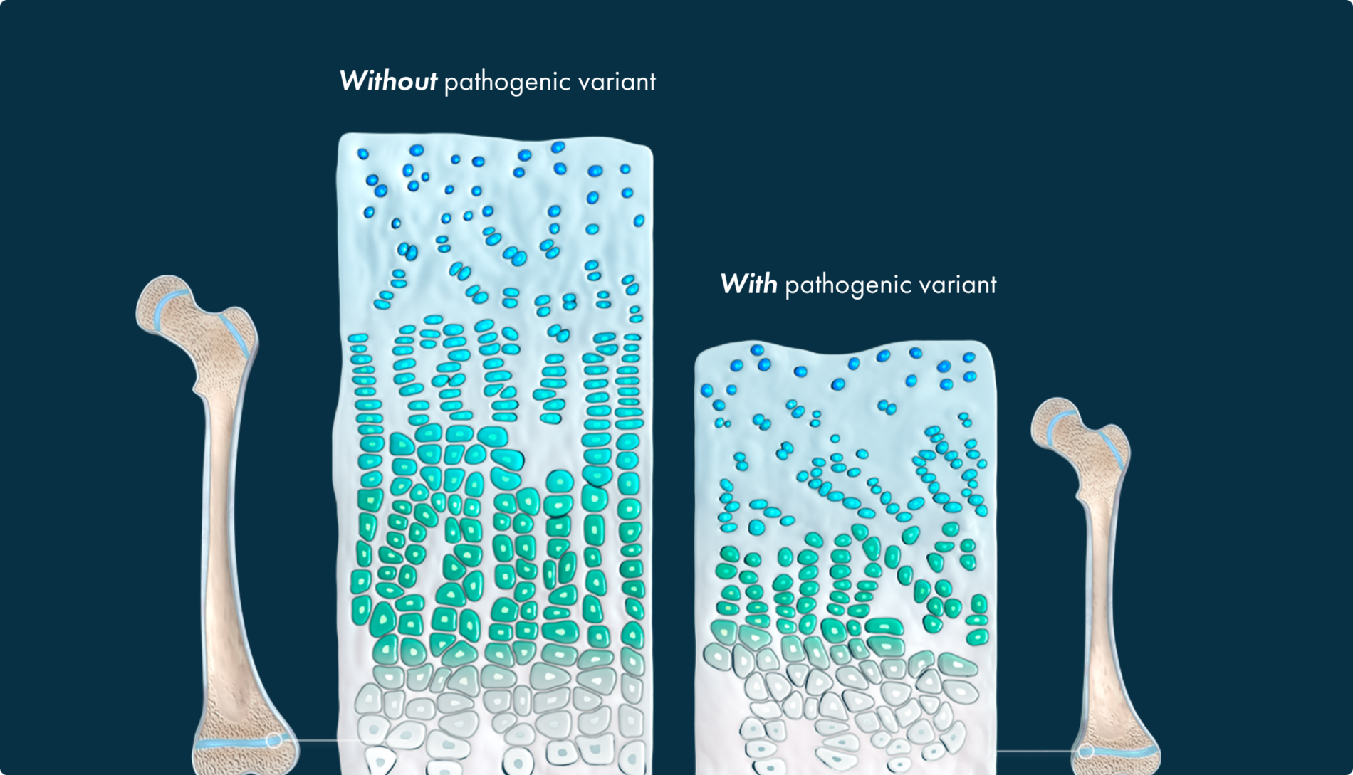
Cartilage growth plate (without pathogenic variant)9,13,14

Cartilage growth plate
  • Cartilage is composed of chondrocytes and extracellular matrix
  • Chondrocytes play a key role in endochondral ossification, directly contributing to the elongation of bones throughout development
Image of cartilage growth plate without achondroplasia
  • Chondrocytes result from the differentiation of embyronic mesenchymal cells
  • Chondrocytes undergo proliferation
Image of cartilage growth plate without achondroplasia
  • Cartilage matrix is secreted
  • Chondrocytes undergo hypertrophy, establishing a new extracellular matrix

Chondrocyte and CNP pathway (without pathogenic variant)15,16

  • Two signaling pathways play an important role in regulating the function of chondrocytes
  • FGFR3 activation signals to slow bone growth
  • CNP (C-type natriuretic peptide) pathway counteracts FGFR3 signals
  • Natriuretic peptide receptor B (NPRB) activation by CNP blocks the FGFR3 signal to sustain bone growth

The gain-of-function pathogenic variant causes FGFR3 to excessively generate signals to slow down bone growth, overwhelming the counteracting signaling from the NPRB/CNP pathway, and resulting in impaired bone growth.4,16

CNP pathway (without pathogenic variant)

CNP pathway (with pathogenic variant)

Cartilage growth plates

This leads to a multisystemic impact that parents may not be prepared for.

Most parents are of average stature, which means they may need your expertise to prepare them for the possible complications caused by impaired bone growth.4,17

Test your knowledge

Achondroplasia occurs in approximately _________ live births worldwide. 

Silhouette of a child with achondroplasia highlighting five key physical characteristics

Achondroplasia complications

Common achondroplasia complications can be expected across an individual’s lifetime.

Resource library

Everything you need to deepen your achondroplasia knowledge

References:

  1. Ireland PJ, Pacey V, Zankl A, Edwards P, Johnston LM, Savarirayan R. Optimal management of complications associated with achondroplasia. Appl Clin Genet. 2014;7:117-125. Published online June 24, 2014.
  2. Wynn J, King TM, Gambello MJ, Waller DK, Hecht JT. Mortality in achondroplasia study: a 42-year follow-up. Am J Med Genet A. 2007;143A:2502-2511.
  3. Waller DK, Correa A, Vo TM, et al. The population-based prevalence of achondroplasia and thanatophoric dysplasia in selected regions of the US. Am J Med Genet A. 2008;146A(18):2385-2389.
  4. Pauli RM. Achondroplasia: a comprehensive clinical review. Orphanet J Rare Dis. 2019;14(1):1.
  5. Laederich MB, Horton WA. Achondroplasia: pathogenesis and implications for future treatment. Curr Opin Pediatr. 2010;22(4):516-523.
  6. Hoover-Fong J, Scott CI, Jones MC; Committee on Genetics. Health supervision for people with achondroplasia. Pediatrics. 2020;145(6):e20201010.
  7. Chilbule SK, Dutt V, Madjhuri V. Limb lengthening in achondroplasia. Indian J Orthop. 2016;50(4):397-405.
  8. Hoover-Fong J, Schulze KJ, McGready J, Barnes H, Scott CI. Age-appropriate body mass index in children with achondroplasia: interpretation in relation to indexes of height. Am J Clin Nutr. 2008;88:364-371.
  9. Matsushita T, Wilcox WR, Chan YY, et al. FGFR3 promotes synchondrosis closure and fusion of ossification centers through the MAPK pathway. Hum Mol Genet. 2009;18(2):227-240.
  10. Berendsen AD, Olsen BR. Bone development. Bone. 2015;80:14-18.
  11. Clarke B. Normal bone anatomy and physiology. Clin J Am Soc Nephrol. 2008;3(Suppl 3):S131-S139.
  12. Hill MA. Musculoskeletal system – bone development timeline. Embryology. June 19, 2020. Accessed September 4, 2020. https://embryology.med.unsw.edu.au/embryology/index.php/Musculoskeletal_System_-_Bone_Development_Timeline.
  13. Xie Y, Zhou S, Chen H, Du X, Chen L. Recent research on the growth plate: advances in fibroblast growth factor signaling in growth plate development and disorders. J Mol Endocrinol. 2014;53(1):T11-T34.
  14. Mackie EJ, Tatarczuch L, Mirams M. The skeleton: a multi-functional complex organ: the growth plate chondrocyte and endochondral ossification. J Endocrinol. 2011;211(2):109-121.
  15. Horton WA, Hall JG, Hecht JT. Achondroplasia. Lancet. 2007;370(9582):162-172.
  16. Vasques GA, Arnhold IJ, Jorge AA. Role of the natriuretic peptide system in normal growth and growth disorders. Horm Res Paediatr. 2014;82(4):222-229.
  17. Hecht JT, Bodensteiner JB, Butler IJ. Neurologic manifestations of achondroplasia. Handb Clin Neurol. 2014;119:551-563.